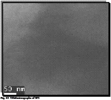
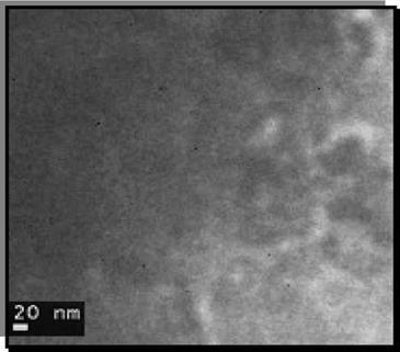
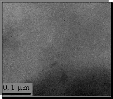
TEM complements XRD by observing a very small section of the material for the possibility of intercalation or exfoliation. It also provides information about the particle size and nanodispersion of particles. It however, supplies information on a very local scale. However, it is a valuable tool because it enables us to see the polymer and the filler on a nanometer scale.
The images, in Fig.12, 1 3 and Fig. 14, provide information regarding likely sizes of particles present in the matrix. Figure shows each of the polymer nanocomposite systems with critical loading of modified red mud. In Fig. 12 the silicate galleries of the organically modified red mud showed partial exfoliation and intercalation as depicted by the ridges in the image. While SP5 and PRM4 showed completely intercalated system. Nanoparticles showed agglomeration in some parts of the composite films due to the conformation of the polymer chains adhered to the nanoparticles.
Particle sizes on the TEM images are worth noting. In Figure 12, several ORM particles in PVA are ~8 nm, in Figure 13, BRM in PVA are as small as ~12 nm, and in Fig. 14, PRM in PVA ranges to a size of ~23 nm. Thus, it could be inferred, that there were numerous platelets that were expanded by the penetration of polymer chains.
|
|
|
|


