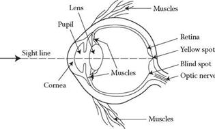
Figure 10.5 shows a cross-section of a human eye. The incoming light is focused by the lens and falls onto the retina that lines the back of the eye. Different parts of the retina have different types of light-sensitive cells on them. In the central section
|
TABLE 10.5 Coloured Light on Different Coloured Surfaces
Source: Modified from Woodson (1981). |
|
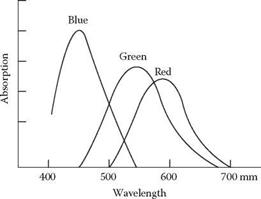
FIGURE 10.4 Relative sensitivity of the eye for different wavelengths of light. |
|
FIGURE 10.5 Cross-section of the human eye. |
(fovea) are cones (see Figure 10.6), which are less sensitive to light. The cones are used for detailed imaging, and have a very different ability to distinguish detail from the rods that are found more peripherally around the retina; the rods are considerably more sensitive to light. The rods are used mainly for night vision and the cones for daylight vision. The cones help us distinguish colour.


