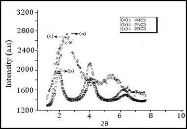
The wide angle diffraction pattern of PRC2 and PRC3 films are shown in Fig. 5. The crystallinity of the composite membranes is mainly due to Phe. The characteristic diffraction peak for PRC2 and PRC3 were observed at a 20 value of 2.36o and 2.26o. The diffraction peak at 20 = 2.26 o is considerably broadened and the interplanner distance has also widened compared to the raw red mud and PRC2 nanocomposite, indicating intercalation of the phenoxy matrix within the nanofiller of the nanocomposite membranes.
The polymer nanocomposite films showed decrease in the intensity of the crystalline peak as the loading percentage of organically modified red mud increased. The decrease in the intensity of crystalline peaks showed an increase in the disorderness of the interlayers of modified red mud.
|
Fig. 24. XRD pattern of (a) PRC2 (b) PNC3 (c) PRC3 nanocomposites |
From table 14, it is clear that the basal spacing for PRC3 is larger than the other PRM based nanocomposites materials, thereby giving evidence of better intercalation of polymer matrix within the silicate galleries of filler in case of PRC3 than the rest of nanocomposites.
|
Sample |
20 |
d-spacing (Ao) |
|
PRC2 |
2.36 |
37.56 |
|
PRC3 |
2.26 |
39.48 |
|
PNC3 |
2.11 |
41.62 |
|
Table 14. XRD results of Phe-PRM and Phe-ORM based nanocomposite membranes |
The X-ray diffraction pattern of phenoxy-organically modified red mud nanocomposite membranes are shown in fig. 24.The characteristic diffraction band of PNC3 was observed at 2 0 = 2.11o. The increase in the interlayer distance of silicate galleries of the organically modified red mud indicated that Phe has successfully intercalated into the silicate layers.


